
Canine in palate
Definition
These are those canines of the upper dental arch that do not erupt when they should, around the age of eleven, and in their place remain the milk canines. It is not infrequent that the lack of the definitive canine in the mouth goes unnoticed by the patient until the milk canine begins to move and finally falls out, in adulthood. The frequency of canine included in the palate -CIP-in the general population is around 1.7%.
Causes
The lack of space in the arch or the persistence of the deciduous canine, usually pointed out as the cause of the inclusion of the canine, are not relevant. On the one hand, people with CIP do not usually present significant space limitations in the arch and, on the other hand, the persistence of the deciduous canine is the consequence, and not the cause, of the ectopic eruption -out of place- of the permanent canine. The genetic origin of CIPs is the most probable etiology in most of them. They go in this direction:
- The high family predisposition of the CIPs.
- Its frequent association with other dental anomalies (agenesis -absence of permanent teeth-, small teeth, other ectopic teeth, delayed eruption).
- The difference in frequency between sexes – females more than males.
- The high frequency of bilaterality, as well as racial differences.
Diagnosis
Clinically: we will suspect the existence of CIP:
- In adults, due to the persistence of the baby tooth -very different from the permanent canine in size and enamel characteristics- and/or the absence of the primary and permanent canine.
- The palpation of them in children is asymmetric (only one is palpated).
- Do not palpate in patients older than 11 years.
- They are palpated on the palate.
- When there is displacement of the lateral incisor in the absence of the canine on the same side.
Radiologically:
If a canine is not palpable, and by dental age it should be in the arch, the next step is to find out where it is (vestibular – outside – or palatal), and how it is (what is its three-dimensional disposition: more or less horizontal-medial-cranial), as well as its relationship with the roots of the neighboring teeth, especially the lateral incisor.
Orthopantomography or panoramic radiography usually gives us enough information to determine whether the canine is palatally or vestibularly located. Nowadays, we recommend routinely requesting a CAT scan-computed axial tomography-to know its precise three-dimensional location, as well as its relationship with the roots of the neighboring teeth, which is very useful information to “discover” it during surgery.
What to do?
In the case of an IPC we can choose to (a) do nothing, (b) extract it or (c) bring it orthodontically into the arch – transalveolar autotransplantation is not discussed here.
In children and adolescents, parents understand very well that the best therapeutic option is to place the permanent canine in the dental arch.
In adults, initially, doubts usually arise, generally due to a certain lack of information. Basically they ask themselves whether it is worth placing the tooth included in the arch with orthodontics -time and appliances-, or whether it is better to place a false tooth -prosthesis- in its place. In a very schematic way we will say:
- A natural tooth is always better than a false one.
- That the placement of a false tooth to replace the included canine involves the placement first of an implant -screw in the bone- and then of the false tooth on top of it -crown.
- That the space available in the dental arch is smaller than that necessary to place a permanent canine, so that in case a false tooth is placed, it will also look small.
- That it is not advisable to leave the canine included in the palate indefinitely: although infrequent, some canines move over time and can cause damage to the roots of the anterior teeth -incisors-, so it is advisable to extract them.
For all of the above reasons, the best option, in the vast majority of cases, will be to place the included canine in its place with orthodontics .
Exceptionally, it will be recommended to extract the CIP:
- When its location on the palate is very unfavorable.
- When there is root resorption – wear of the roots – of the neighboring teeth – depending on the magnitude and viability of the same – and their orthodontic displacement may aggravate them.
- When the first premolar – the next tooth in position in the arch – occupies or can occupy its place in the arch with orthodontics, fulfilling the function of the canine acceptably.
- In cases where extensive prosthetic rehabilitation is to be performed;
- When it presents anatomical anomalies – anomalous shape.
- When ankylosed – it will only be discovered in the first weeks of CIP traction, after surgical exposure. Its frequency increases with age.
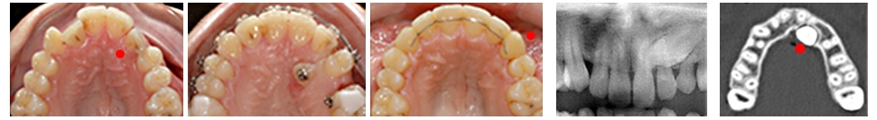
Treatment Sequence: once placed on the palate
- Brackets are placed in order to create the necessary space in the arch and to use the whole arch as an anchorage unit from which the canine can be tractioned.
- Surgical exposure is performed.
- The canine is progressively tractioned into place in the arch.
In adults, treatment begins with the placement of brackets on all the teeth of the upper arch. The baby canine is kept in the arch, for esthetic reasons, until the canine begins to be tractioned and its absence is concealed. In adolescents we will begin treatment when the patient has the definitive dentition, with or without the presence of the molars at the age of 12. If the patient presents other types of dental or skeletal problems, depending on their nature, they will be treated before or during the treatment of the included canine. Sometimes, when the CIP is palpated, we perform the surgical exposure of the canine before placing the appliances. Appliance: brackets on all teeth in the arch. We do not use anchorage braces. The appliances used are basically the same as in a simple tooth alignment case. Surgery: it is an outpatient procedure, not traumatic, performed under local anesthesia. A window is made on the mucosa of the palate to access the enamel of the canine. During this procedure a pin is glued on the canine to be able to pull the canine – similar to a bracket. Duration: the placement of a CIP in your arch, in the absence of other accompanying alterations, usually requires about 24-28 months of treatment.