
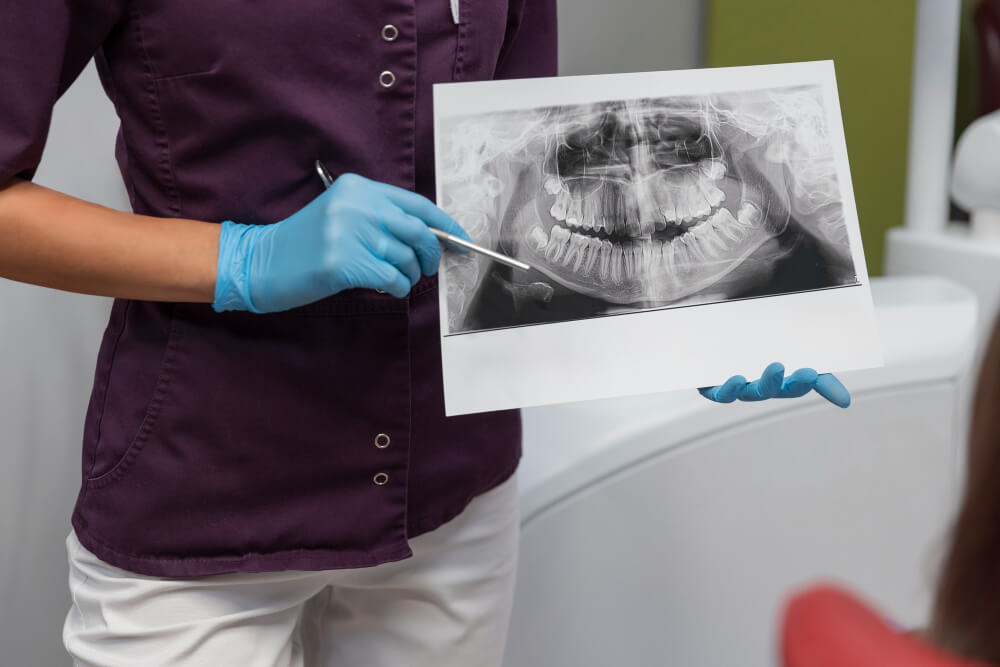
Orthopantomography, also known as panoramic dental radiography, is a radiological image that shows an overview of the mouth, including the teeth, jaw, maxilla and other adjacent structures, such as the paranasal sinuses and temporomandibular joint. It is a fundamental tool in dentistry to evaluate oral health in a complete and quick way.
How is it done?
- Preparation of the patient: The patient should remove any metal objects on the head or neck (such as earrings, necklaces or piercings) that may interfere with the image.
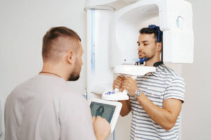
- Positioning: The patient stands in front of the X-ray machine. He/she is asked to bite down on a small plastic piece and to keep his/her head in a specific position so that the image comes out as clear as possible.
- Image acquisition: The equipment performs a semicircular movement around the patient’s head, capturing a panoramic image of the mouth and jaw. The acquisition is fast and non-invasive, lasting only a few seconds.
What is it for?
Orthopantomography has multiple uses in dental and maxillofacial practice, among which the following stand out:
- Caries detection: Allows the identification of caries that may not be visible in a basic clinical examination.
- Evaluation of impacted teeth: It is useful to locate teeth that have not erupted, such as wisdom teeth.
- Diagnosis of periodontal diseases: Helps evaluate the health of the bone around the teeth, which is key in the diagnosis of periodontitis.
- Treatment planning: Used for planning orthodontics, implants and other dental treatments by providing a complete view of the bone structure.
- Detection of lesions or anomalies: such as cysts, tumors or fractures in the jaw.
Orthopantomography is essential in modern dentistry because it allows a complete and accurate view of the oral area, facilitating effective diagnosis and treatment.
Is orthopantomography used in Orthodontics?
Yes, orthopantomography is widely used in orthodontics. It is essential for the diagnosis, planning and follow-up of orthodontic treatments, as it provides a detailed panoramic image of the mouth and bone structures.
How is it used in orthodontics?
In orthodontics, orthopantomography helps to:
- Evaluate the general condition of the teeth and bone: Allows the orthodontist to see how the teeth are aligned and if there are any problems in the bone, such as bone loss or malformations.
- Diagnose retained or supernumerary teeth: It is common for some teeth not to erupt (such as canines), or for extra (supernumerary) teeth to exist, and orthopantomography helps to identify them and plan their management.
- Visualize wisdom teeth (third molars): The position of wisdom teeth is important in orthodontics, as they can affect the alignment of the other teeth. Orthopantomography makes it possible to evaluate them and decide if they need to be extracted.
- Planning tooth movement: In orthodontic treatment, the orthodontist must consider the size, shape and position of the roots of the teeth. Orthopantomography shows these characteristics, helping to plan safe and effective tooth movement.
- Evaluate bone structure: By showing the maxillary and mandibular bone, orthopantomography helps evaluate bone growth and development, which is crucial in the treatment of young or growing patients.
Orthopantomography is an essentialtool in invisible orthodontics because it provides a comprehensive view of the mouth and allows the orthodontist to plan and monitor treatment effectively and safely.
